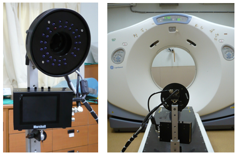
Respiratory Gating
Respiratory gating is a technique used in medical imaging, particularly in radiation therapy and diagnostic imaging such as PET (Positron Emission Tomography) and MRI (Magnetic Resonance Imaging). It’s employed primarily to account for the motion of organs affected by respiration, mainly the lungs and sometimes the liver.
Here’s how it works:
Respiratory Monitoring: The patient’s respiratory motion is monitored using various methods such as a bellows system, external markers, or internal markers like fiducial markers implanted near the organ of interest.
Signal Acquisition: The monitoring system captures the respiratory signal, typically in the form of a waveform or a series of data points that represent the phase of the respiratory cycle.

Data Processing: The acquired respiratory signal is analyzed to identify the different phases of the respiratory cycle, such as inhalation and exhalation.
Gating: During imaging, such as during a CT scan or MRI, the acquisition of image data is synchronized with the respiratory cycle. Imaging is triggered to occur only during specific phases of the respiratory cycle, typically during end-expiration (when the lungs are fully deflated and motion is minimal). This gating ensures that the images are acquired when the organ of interest is in a relatively stable position, reducing motion artifacts and improving image quality.
Image Reconstruction: The acquired data is reconstructed into images, taking into account the gating information. This may involve sorting the acquired data into different respiratory phases and reconstructing images separately for each phase.
Clinical Applications: Respiratory gating is particularly useful in radiation therapy, where it helps in accurately targeting tumors in organs affected by respiratory motion, such as lung tumors. It also finds applications in diagnostic imaging to improve the visualization of organs affected by respiratory motion, enabling more accurate diagnosis and treatment planning.
Overall, respiratory gating enhances the quality and accuracy of medical imaging by accounting for the inherent motion of organs due to respiration, thereby improving the effectiveness of both diagnosis and treatment.
